


CT-SCAN Laboratory
* EQUIPTMENT:

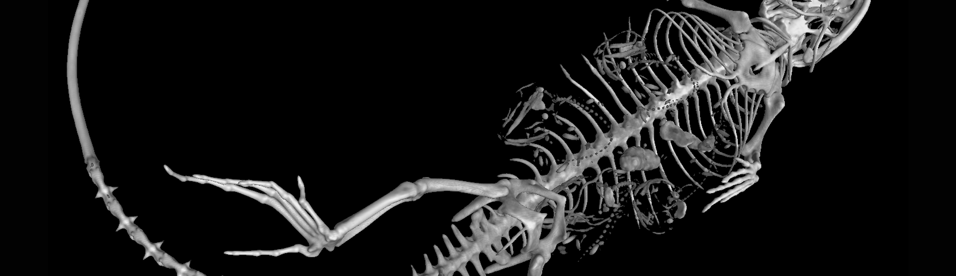
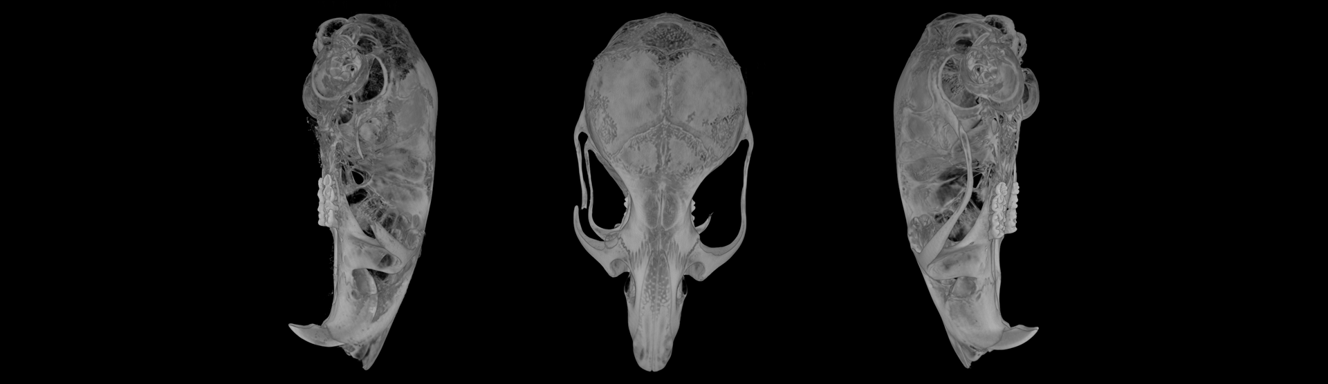
The Non-Destructive Techniques Service has an X-ray computerized tomography machine, NIKON CT-SCAN-XT H-160, with software for data evaluation. The equipment allows the analysis of variables such as areas, volumes, porosity, thickness, density, etc., all without destroying the sample. It covers a very wide range of applications, including the inspection of plastic parts, complex mechanisms, as well as the analysis of materials and natural specimens, fossils, gems...
The parameters of the X-ray tube that it has incorporated and the four targets available to the laboratory allow the selection of the best configuration according to the object being analyzed; rock, bone, tooth, soft tissue, etc. It can reach a maximum power of 160 kV. The MicroCT allows samples to be scanned from a few millimeters in size to 6.5-7 cm. Thanks to the equipment's high-precision microfocus, resolutions up to 3 microns can be achieved (only for very small samples). The maximum allowed weight for the piece of study is 15 Kg.
* SESSIONS
The sessions will always be assisted by a specialist technician.
For the correct functioning of the Service, users will first contact the laboratory through the contact form or by email. After that, if necessary, they will contact the specialist by telephone in order to determine free dates and the duration of the agreed work.
* CONDITIONS OF DELIVERY OF SAMPLES AND RESULTS
The samples will be delivered to the Service either prior to the reserved session or on the same day of the session, always respecting the punctuality of the assigned time. Samples can also be sent by mail, as long as they are received prior to the day of the assigned session.
The laboratory will contact the user once the tomographies have been performed to agree on the gathering of the samples and the delivery of the results, as well as the format of the same.
If the user wishes, the Service can provide instructions to download a 3D viewer for free, which will allow them to view their scans.
It is necessary to come with an external hard disk or pen drive to take the results.
* PREPARATION OF SAMPLES FOR THE SESSION
Computed tomography samples do not require any preparation, although it is highly recommended to consult with the specialist technician if they are very delicate. In addition, and depending on the type of sample, the possible prior assembly of the samples by the user will also be discussed with the technician.
* PRINCIPLES OF THE TECHNIQUE
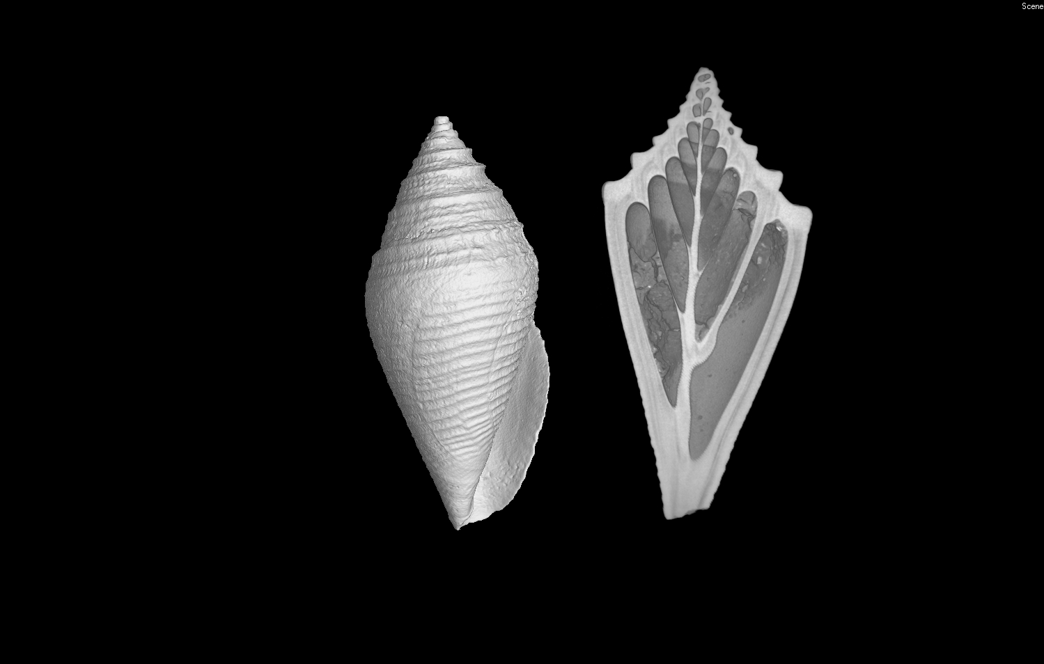
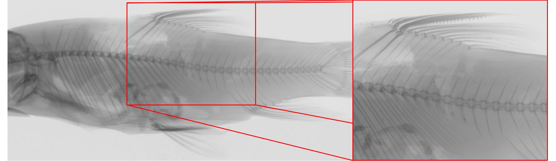
X-ray computed tomography fundaments are simple: an object is placed on a rotating stand between the X-ray source and the detector. A high-precision microfocus generates the radiation at the source, and the X-rays generated go through the sample as it rotates 360º. The digital detector captures a sequential series of 2D images of the X-ray patterns going through the object, showing different shades of gray depending on the material and the geometry. Dense materials such as iron, copper or lead are the darkest areas, while thinner or lighter materials such as plastic, paper or air are the lighter areas.
The 2D images of the tomography then go to a software that, by means of a reconstruction algorithm, provides a 3D volumetric map of the object, thanks to which both the interior and the exterior of the sample can be observed.
Staff
Cristina Paradela Guerrero
Marta Furió Vega
Laura Tormo Cifuentes