


Scanning Electron Microscopy Laboratory
* EQUIPMENT:

- Scanning Electron Microscope FEI INSPECT. This microscope operates in two vacuum modes (high and low vacuum) with secondary and backscattered electron detectors for both vacuum modes.
This microscope has an OXFORD INSTRUMENTS analytical-INCA X-ray energy dispersive analysis system and a CATHODOLUMINESCENCE detector (model GATAN MonoCL3) with a cooling system with liquid nitrogen and spectral magnification up to the infrared.

- Scanning Electron Microscope FEI QUANTA 200. This microscope operates in three vacuum modes (high vacuum, low vacuum and enviroment mode) with secondary and backscattered electron detectors for all vacuum modes.
The microscope has an integrated OXFORD INSTRUMENTS Analytical-INCA analysis system with two X-ray detectors that can be used simultaneously and alternatively: one EDS (Energy Dispersive Spectroscopy) and the other WDS (Wavelength Dispersive Spectroscopy).
SESSIONS
In both microscopes the sessions are always WITH technical assistance.
The laboratory technicians try to adjust the schedule and the needs of the different working groups. For this, sessions of different length of time and schedules have been prepared to add more versatility and adjustment to the needs of the users with the laboratory’s agenda.
* CONDITIONS OF DELIVERY OF SAMPLES AND RESULTS
Since the users are normally present in the laboratory (see the covid-19 action plan of the analysis Service by Non-Destructive Techniques), at the end of the session they will take their samples and the results obtained. When the user requests a routine session that the technicians can carry out without his presence, and always in agreement with him, the results, the report and the samples will be sent to him.
It is highly recommended to always come with an external hard drive or pen drive to take the results at the moment.
* PREPARATION OF SAMPLES FOR THE SESSION
♦ INSPECT AND QUANTA 200 IN HIGH VACUUM. It depends on the type of sample, but they must be dry solids, or in the last phase of dehydration, with the fixation and post-fixation treatments already carried out. When the samples require critical point drying, it is necessary to reserve an appointment for it at least one week before their observation in SEM. Wet samples, which require a critical point, will be delivered to the service in a solution of 100% acetone or absolute alcohol.
♦ INSPECT AND QUANTA 200 IN LOW VACUUM AND ENVIRONMENTAL. The samples can be put in without prior preparation, without coating or dehydrating.
* PRINCIPLES OF THE TECHNIQUE
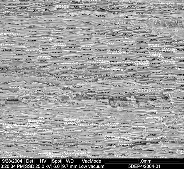
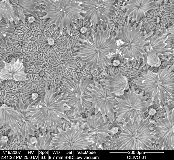
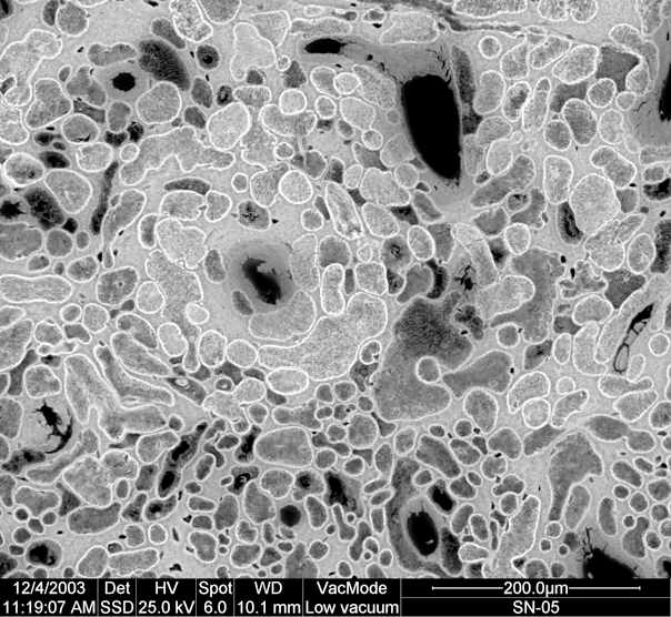
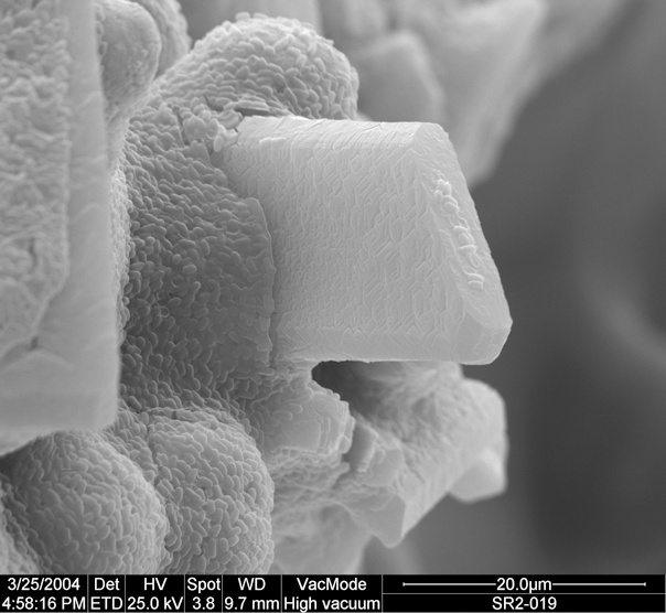
The scanning electron microscope is an instrument that allows the observation and characterization of inorganic and organic materials studying their surfaces. From the jet of electrons that falls on the sample, different types of signal are generated from the sample (secondary electrons, backscattered, X-rays, etc.) that are used to examine many of its characteristics (topography or density of the different components).
The X-ray Energy Dispersion (EDS) analyzer is a non-destructive analysis technique where the radiation used is the characteristic X-rays emitted by the sample as a result of electron bombardment. The analysis of this radiation provides analytical information on the composition of the total sample or areas of it up to a few microns in diameter. These analyses can be performed on non-coated biological samples by providing a percentage elemental composition spectrum. Results in non-coated biological samples are satisfactory. However, more conclusive values are attained if the samples are polished and if the biological samples are previously coated. Both the QUANTA 200 and the Inspect are equipped with EDS detectors.
The X-ray wavelength dispersion (WDS) analyzer installed in the QUANTA 200 is a non-destructive analysis technique where it is possible to analyze elements present in the sample in very low concentration (non-detectable by EDS and more precise) thus complementing the Energy Dispersive analysis. These analyses can also be carried out on biological samples, although the results are of better quality when the samples are flat, further improving the results in polished and coated samples.
The Catodoluminescence (CL) analyzer is a non-destructive analysis technique that makes a spectral analysis of the luminescence that the sample has and emits when bombarded by electrons, providing analytical and structural information about the sample. The GATAN MonoCL3 cathodoluminescence detector of the MNCN in combination with the SEM Inspect, in which it is installed, have a high resolution capacity and very good quantum efficiency, which allows obtaining spectra even in samples with very low emissions, such as hydroxyapatites, carbons, woods, kidney stones... It is a complementary technique to RAMAN since samples that have luminescence and cannot be seen by Raman spectroscopy can be analyzed with the Catodoluminescence detector and thus be able to analyze the molecular structure in the visible light spectrum and infrared.
Schedule
De 8:30 a 17:00
Staff
Laura Tormo Cifuentes
Marta Furió Vega
Pedro Valverde Lobato
Manuel Linares Ruiz